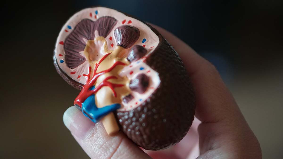
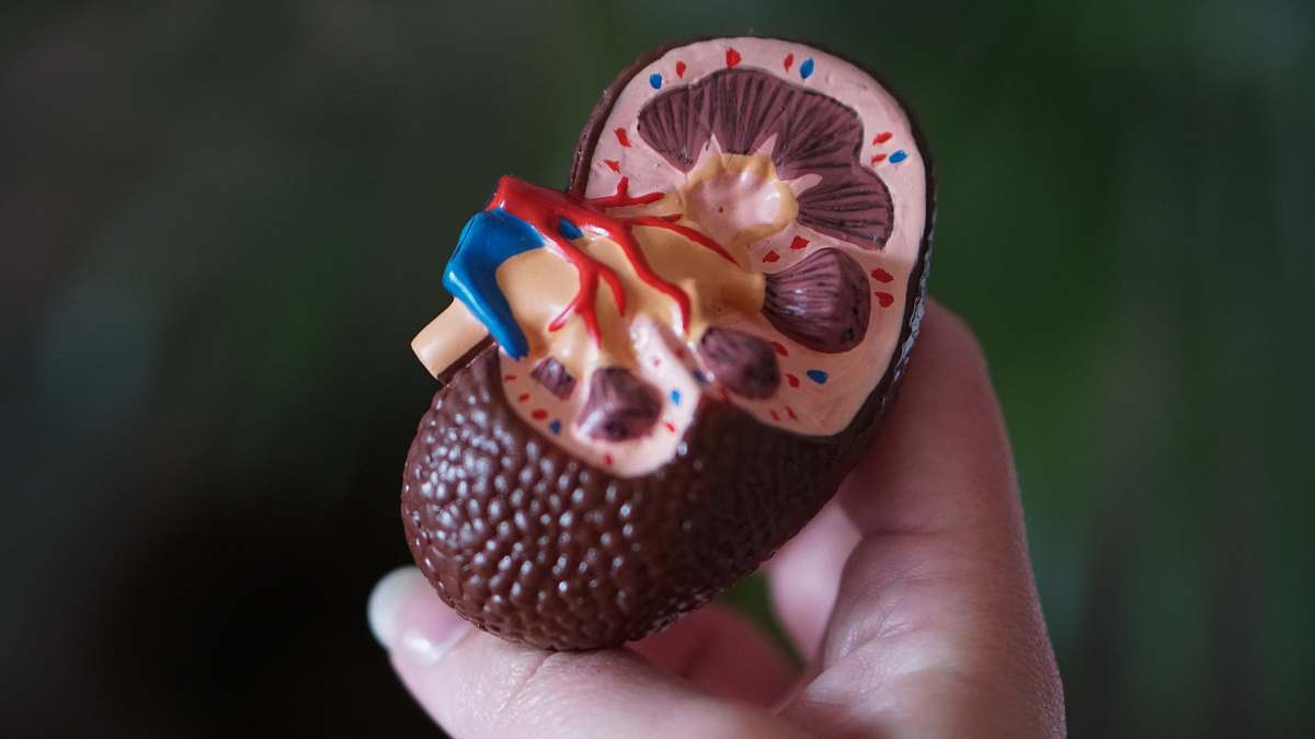
Microscopic Section of Kidney Tissue
High Resolution JPEG Picture of science medical anthropotomy physiology microscopic section of kidney tissue background
This image showcases a highly detailed microscopic section of kidney tissue. The specimen highlights the intricate structures of nephrons and surrounding tissue, providing valuable insight into renal physiology. The clarity and precision of the capture make it suitable for scientific analysis and educational purposes. This visual representation serves as a critical tool for understanding kidney functions and pathology.
This image can be utilized in various contexts, including medical textbooks, research articles, and educational workshops. It is particularly useful for health professionals and students learning about renal anatomy and diseases. Additionally, the image can be featured in informative blogs, medical presentations, and online courses related to nephrology. It can also enhance infographics, social media posts, and educational eBooks focused on health sciences.