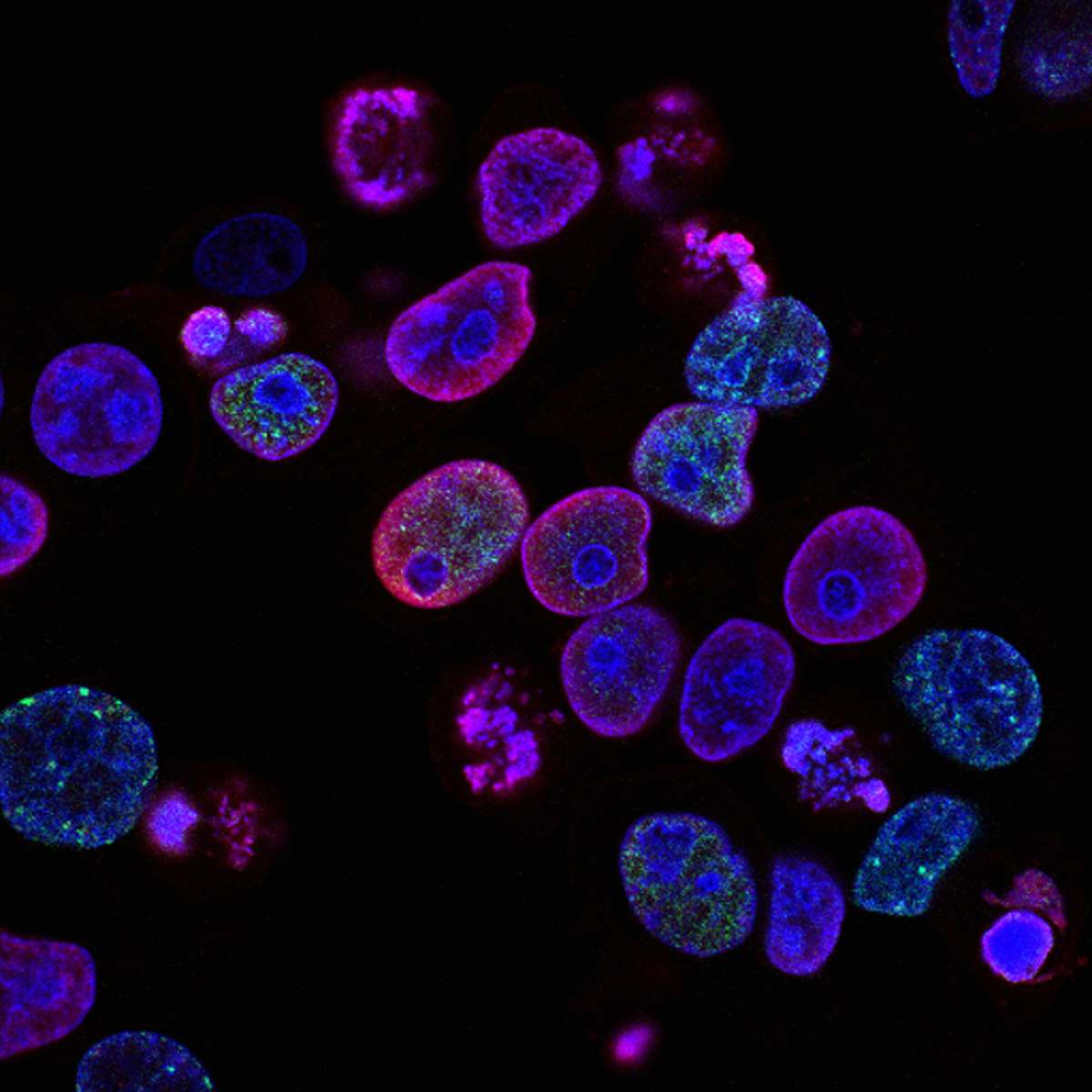
Monkey Pox Cells Under Microscope
High Resolution JPEG Picture of Monkeypox Virus. 3D Render
This image showcases monkey pox cells viewed through a microscope slide, highlighting their unique structure and characteristics. The high-resolution capture allows for detailed examination of the cells, making it a valuable resource for research and educational purposes. The clear depiction aids in understanding the morphology of the virus, crucial for biology and medical studies. The design suits both academic and professional contexts.
This image can be effectively utilized in various settings, including academic presentations, health brochures, and medical textbooks. Researchers can incorporate it into studies or reports on viral diseases, while educators may use it in lesson plans or digital content. Additionally, health-related blogs and social media platforms can leverage this image to enhance visual appeal and provide informative content on viral infections.